
Figure 2.
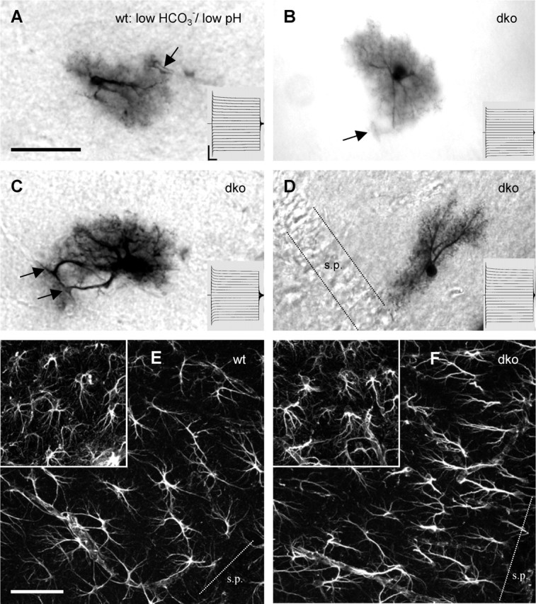
dko astrocytes and wt astrocytes share similar morphology and whole-cell currents. A, Tracer-filled astrocyte in a wt hippocampal slice. Low bicarbonate bath solution, pH 6.4, was applied 30 min before and during whole-cell recordings (20 min) to block gap junctional coupling. Under these conditions, this cell was tracer coupled only with two other astrocytes. Note the prominent primary processes and the dense net of fine processes. The arrow indicates a blood vessel encircled by an astrocytic end foot. Scale bar, 50 μm. The inset represents current responses elicited by 50 ms voltage steps from −180 to 0 mV (Vhold of −90 mV). Calibration: 10 ms, 4 nA. B–D represent examples of tracer-filled astrocytes in slices of dko mice, using standard ACSF. Arrows denote astrocytic pericapillary end feet. Insets and calibration as in A. Note that the astrocyte in D displays a bipolar morphology with an orientation perpendicular to the stratum pyramidale (s.p.). E and F depict immunofluorescent anti-GFAP stainings of the stratum radiatum of wt and dko mice, respectively. The insets show stainings of the stratum lacunosum moleculare. Densities of GFAP-positive cells were similar in wt and dko mice, each with a higher density in stratum lacunosum moleculare compared with the stratum radiatum. Note that major processes of stratum radiatum astrocytes often were oriented perpendicularly to the stratum pyramidale. Scale bar, 50 μm.