
Figure 3.
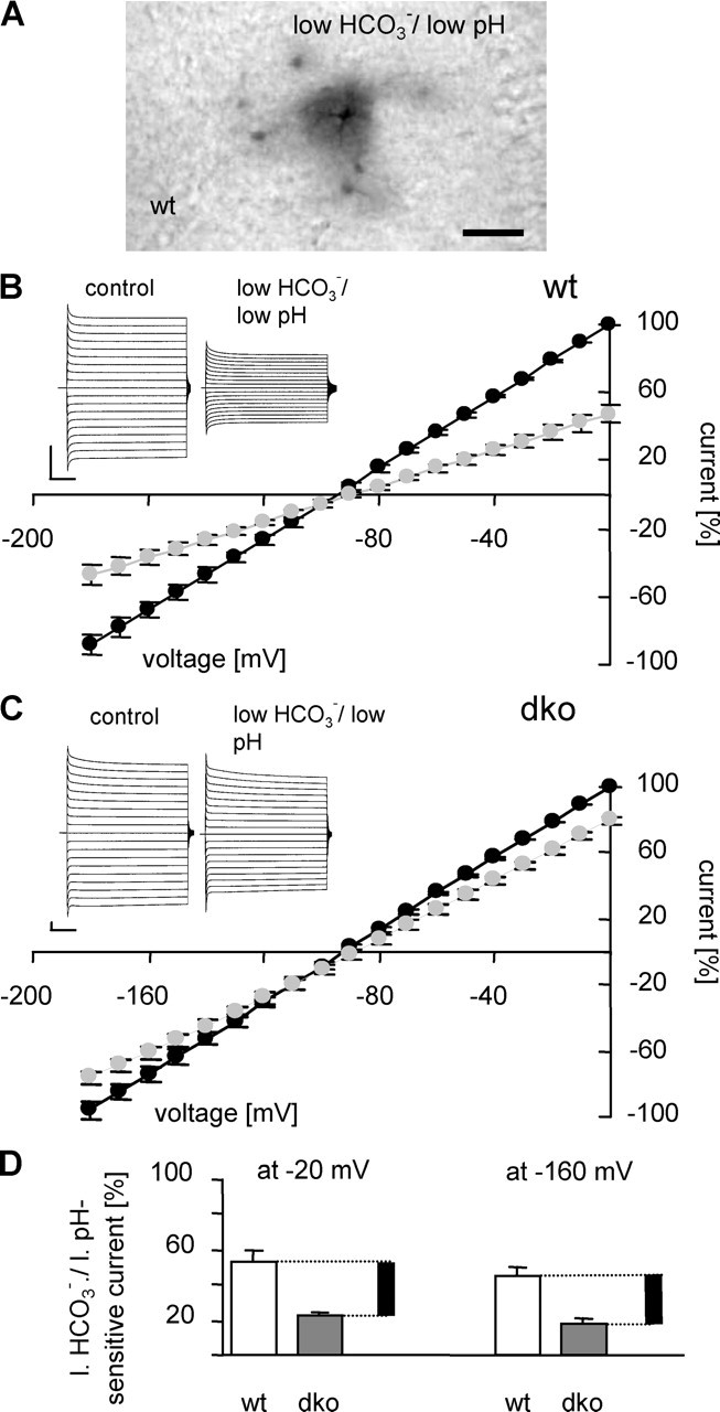
Gap junctions account for ∼30% of whole-cell currents in wt astrocytes. A illustrates the blocking action of l.b./l.pH bath solution, pH 6.4, on tracer coupling in a wt hippocampal slice (30 min preapplication, 20 min recordings). Scale bar, 50 μm. B and C demonstrate the effect of l.b./l.pH solution on whole-cell currents in wt and dko astrocytes, respectively. The insets show representative examples (voltage steps as in Fig. 2); current–voltage relationships in control ACSF (black circles) and l.b./l.pH solution (gray circles) represent means and SEM of five cells. Current amplitudes were normalized to the amplitude recorded at 0 mV. Calibration: 1 nA, 10 ms. D, The amount of bicarbonate-sensitive currents in wt (white bars; 53 ± 5% at −20 mV and 45 ± 5% at −160 mV; n = 5) and dko (dark gray bars; 22 ± 3% at −20 mV and 18 ± 3% at −160 mV; n = 5) mice. For every holding potential, current amplitude in standard ACSF was considered 100%. Black bars represent the extrapolated amount of gap junction-mediated current in wt astrocytes.