
Figure 8.
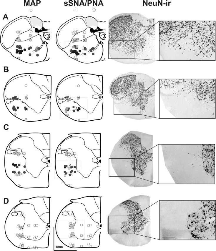
Representative coronal sections at the level of the 14.6 mm caudal to bregma (A), first cervical segment (B), second and third cervical segments (C), and fourth and fifth cervical segments (D) illustrating glutamate injection sites and the evoked changes in MAP (circles), sSNA (squares), and the phrenic nerve (PNA; pentagons). The symbols are graded from dark to light indicating maximal and minimal responses. Black indicates >40 mmHg change in MAP, >100% increase in sSNA, and >60% decrease in the PNA. The letter A indicates sites where the apnea was evoked. Dark gray indicates a response between 20 and 50 mmHg, between 40 and 100% increase in sSNA, and between 30 and 60% decrease in the PNA, whereas light gray indicates a response between 10 and 20 mmHg, between 10 and 20% increase in sSNA, and between 10 and 30% decrease in the PNA. Open symbols indicate no response. The right column shows anatomically matched sections immunohistochemically labeled for neuron-specific nuclear protein NeuN. The largest pressor and sympathoexcitatory responses (100 mm, 50 nl) were found in A, however large responses were also evoked in the region extending into the third cervical segment (B, C). Large responses were not elicited anywhere in the fourth and fifth cervical segments (D). Note that pressor responses were evoked from regions extending into the white matter where scattered neurons were found. ir, Immunoreactive.