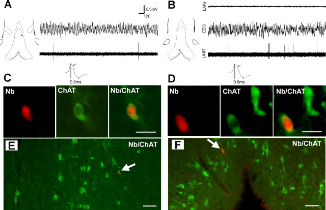
Figure 3.
Discharge properties of two cholinergic neurons recorded in an anesthetized rat (A) and in an unanesthetized rat (B). The red dots on the frontal sections of MS-DB show the location of the neurons. Note the low level of discharge of both units (20 s epochs) in the presence of urethane-induced or wake-related hippocampal theta (EEG). On expended traces, note the hump on the falling phase of the potential (arrow) and long duration of the spike (0.8 ms). C, D, Microphotographs of neurons in A and B. Scale bar, 20 μm. These neurons labeled with neurobiotin (Nb; red) are also immunoreactive for ChAT (green). E, F, Microphotographs at lower magnification showing that the neurons are located in areas with a high density of ChAT+ neurons, i.e., in a dorsoventral band that expands laterally to the midline (A) and in the ventral part of the vDB (B). Scale bar, 50 μm.

