
Figure 4.
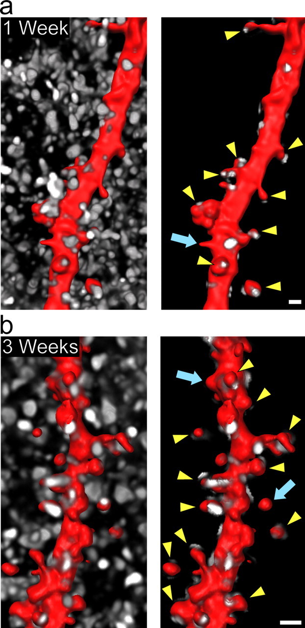
Dendritic spines are innervated in hippocampal slice culture. a, b, SFV PD RFPf-infected slices at 1 and 3 weeks in vitro and immunolabeled for synaptophysin (white) to label presynaptic terminals. At both time points, the majority of CA1 pyramidal cell dendritic spine heads (red) are innervated by presynaptic terminals (arrowheads), whereas only a small proportion are not (arrows). Spines that show association of synaptophysin punctae on the neck are not labeled. Shown are images of the synaptophysin labeling before and after performing a three-dimensional “masking” procedure to preserve only the synaptophysin punctae that are associated with the dendrite (see Materials and Methods). Scale bars, 1 μm.