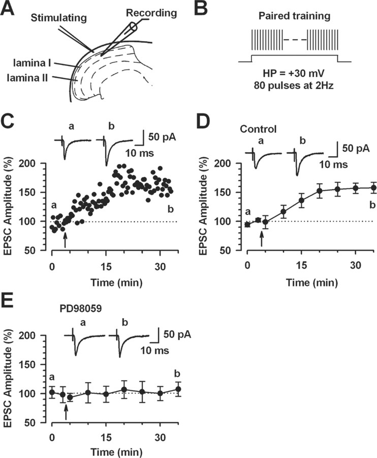
Figure 11.
Activation of Erk for the induction of LTP in the superficial dorsal horn neurons. A, Diagram of a slice showing the placement of a whole-cell patch-clamp recording and a stimulation electrode in the superficial dorsal horn of the spinal cord. B, Schematic illustrating the induction protocol consisting of 80 pulses at 2 Hz while holding at +30 mV (paired training). C, LTP was induced by paired training in the superficial dorsal horn neurons. D, Summary result of the LTP experiments under control conditions (n = 9 neurons). EPSC responses were averaged over 5 min intervals. E, The MEK inhibitor PD98059 (50 μm) in the intracellular solution completely blocked LTP induction (n = 7 neurons). EPSC responses were averaged over 5 min intervals. C–E, Traces show averages of six EPSCs 3 min before (a) and 25 min after (b) the paired training (arrow). The dashed line indicates the mean basal synaptic responses. Error bars indicate SEM.

