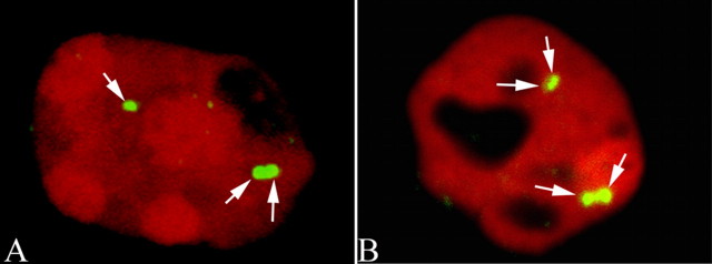
Figure 3.
Confocal images of increased ploidy in some of the neurons of the adult R1.40 transgenic mouse brain. These images were taken with a Zeiss (Thornwood, NY) 510 confocal microscope of neurons in the frontal cortex of a 22-month-old R1.40 mouse. The cell nucleus is revealed with a propidium iodide (red) counterstain. A, B, The hyperploid DNA content of both neurons is illustrated by the three (A, arrows) and four (B, arrows) spots of hybridization of a BAC genomic probe complementary to sequences on mouse chromosome 16. Note that despite the elongated shape of the nucleus shown in A, the three hybridization signals are clearly contained within a single nuclear profile.

