
Figure 5.
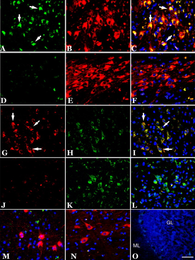
Expression of cell-cycle proteins in subcortical structures in 22-month-old R1.40 mice. A–F, Cyclin A (green) was expressed in TH-positive (red) neurons of the locus ceruleus (A–C, arrows), whereas an aged-matched wild-type control showed only nonspecific cyclin A immunoreactivity (D–F). G–L, Cyclin A (red) was also expressed in the tryptophan hydroxylase-positive (green) neurons of the dorsal raphe (G–I, arrows), whereas an aged-matched wild-type control showed only background cyclin A immunoreactivity (J–L). M–O, No cell-cycle expression was observed in the ChAT-positive (red) neurons of the basal nucleus (M), the TH-positive (red) neurons of the substantia nigra (N), or in the neurons of the cerebellar cortex (O). Cell nuclei counterstained with DAPI (blue) are shown throughout. Scale bars, 10 μm. GL, Granule cell layer; ML, molecular layer.