
Figure 1.
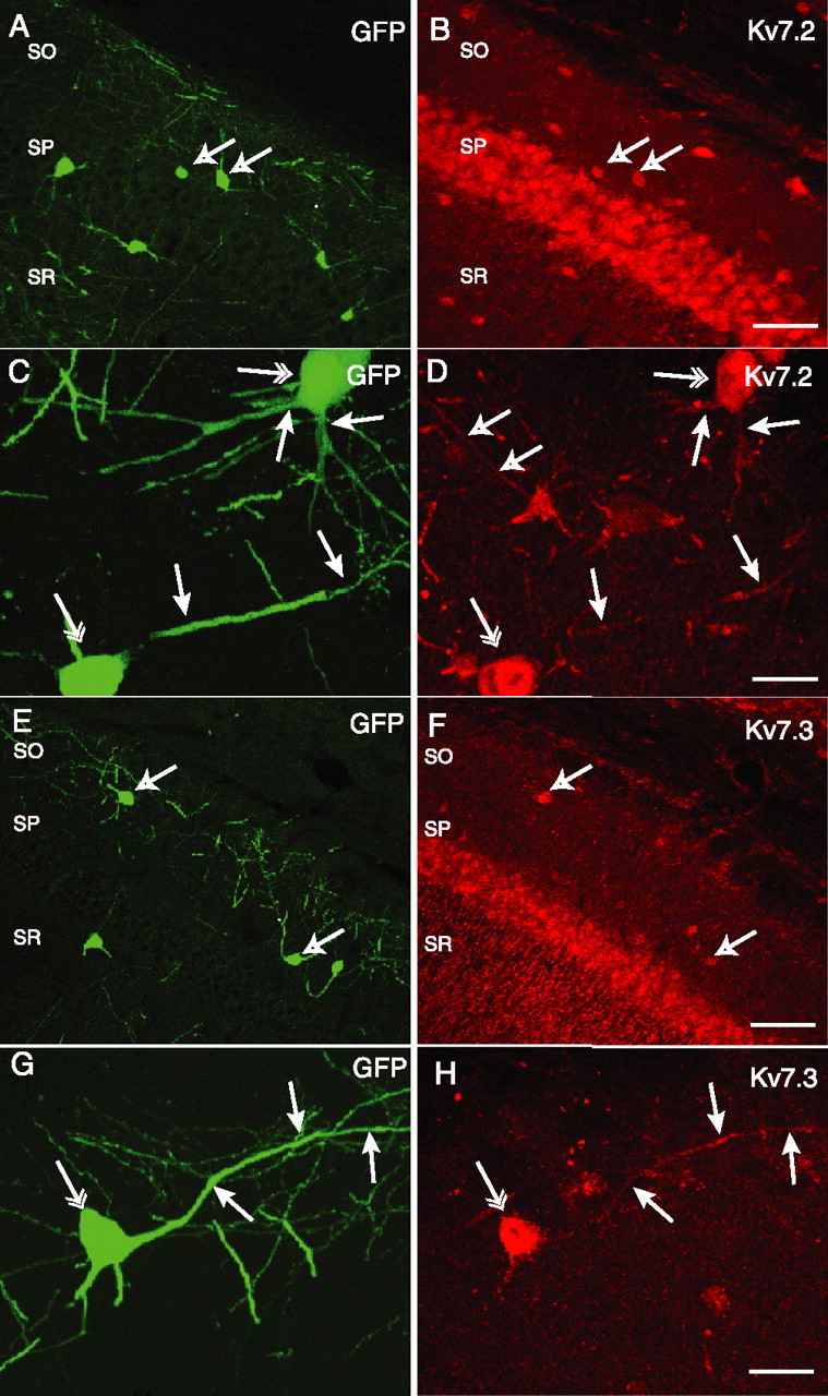
Somatodendritic expression of Kv7.2 (A–D) and Kv7.3 (E–H) in GFP+ hippocampal interneurons. Confocal images of CA1 hippocampal slices from GIN transgenic mice, which express GFP in somatostatin-positive CA1 hippocampal interneurons in SO, SP, and stratum radiatum (SR), are shown. GFP expression (A) and Kv7.2+ immunoreactivity (B) reveal colocalization of Kv7.2 in a subpopulation of GFP+ cells (arrows). GFP+ cells in SO exhibit Kv7.2 immunoreactivity on somata (C, double arrows) and dendrites (C, D, single filled arrows). Open arrows in D indicate a Kv7.2-immunoreactive dendrite from a GFP-negative interneuron. GFP+ interneurons (E) also colocalize with Kv7.3 immunoreactivity (F, arrows). In a GFP+ SO interneuron (G), somatic (H, double arrow) and dendritic (H, single arrows) Kv7.3 immunoreactivity was also apparent. Scale bars: B, F, 100 μm; D, H, 25 μm.