
Figure 1.
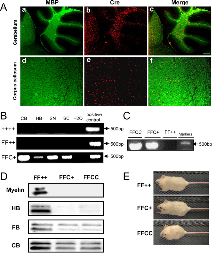
Generation of Fgfr2 conditional knock-out mice. A, Immunolabeling of control cerebellum and corpus callosum with anti-MBP and anti-Cre. Nuclear CNP–Cre expression overlapped with MBP in the cerebellar white matter and corpus callosum, showing that the target cells are oligodendrocytes. Scale bars: a–c, 200 μm; d–f, 100 μm. B, PCR of genomic DNA from wild type (++++), control (FF++; Fgfr2flox/flox,+/+), and FFC+ (Fgfr2flox/flox,cre/+). Primers amplify a 500 bp DNA fragment indicative of the expected deletion in tissue taken from different regions of the CNS and PNS, showing that the Fgfr2 gene sequence has been successfully disrupted in FFC+ mutant mice. CB, Cerebellum; HB, hindbrain; SN, sciatic nerve; SC, spinal cord. C, RT-PCR with primers that identify transcripts with the expected deletion shows that the deleted Fgfr2 mRNA is present in mutant mice with either one (FFC+) or two (FFCC) alleles of cre but not in control mice (FF++). D, Immunoblot analyses of different regions of the brain from mutant mice (FFC+ and FFCC) and control mice (FF++) mice show that the Fgfr2-specific doublet is missing (myelin and myelin-rich hindbrain) or present at only low levels (forebrain and cerebellum, tissues in which other cell types presumably contribute Fgfr2) in the mutant mice. E, At 7 months of age, the body sizes of FFC+ and FF++ mice are comparable, whereas those of FFCC mice are significantly smaller.