
Figure 2.
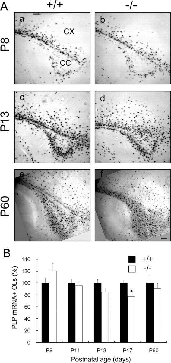
Oligodendrocyte differentiation in Fgfr2 conditional knock-out mouse brain. A, Parasagittal sections from matched regions of forebrains of control (a, c, e) and FFC+ (b, d, f) mice at P8 (a, b), P13 (c, d), and P60 (e, f) were analyzed by in situ hybridization for the OL marker PLP mRNA. PLP mRNA+ cells showed similar patterns of expression in FFC+ (−/−) compared with control (+/+) mice. CX, Cerebral cortex; CC, corpus callosum. Scale bar, 100 μm. B, Quantification of the number of PLP mRNA+ cells that differentiated as a function of time in the corpus callosum of control and FFC+ mice. All PLP mRNA+ cells in the entire corpus callosum region were counted for each section. Four to 10 sections each from four mice from each group and age were analyzed. No significant differences were found between control and FFC+ mice at all ages, except for a slight decrease at P17 (p < 0.05, Student's t test). Black bar, Control (+/+); white bar, FFC+ (−/−). Error bars indicate SEM (n = 4).