
Figure 3.
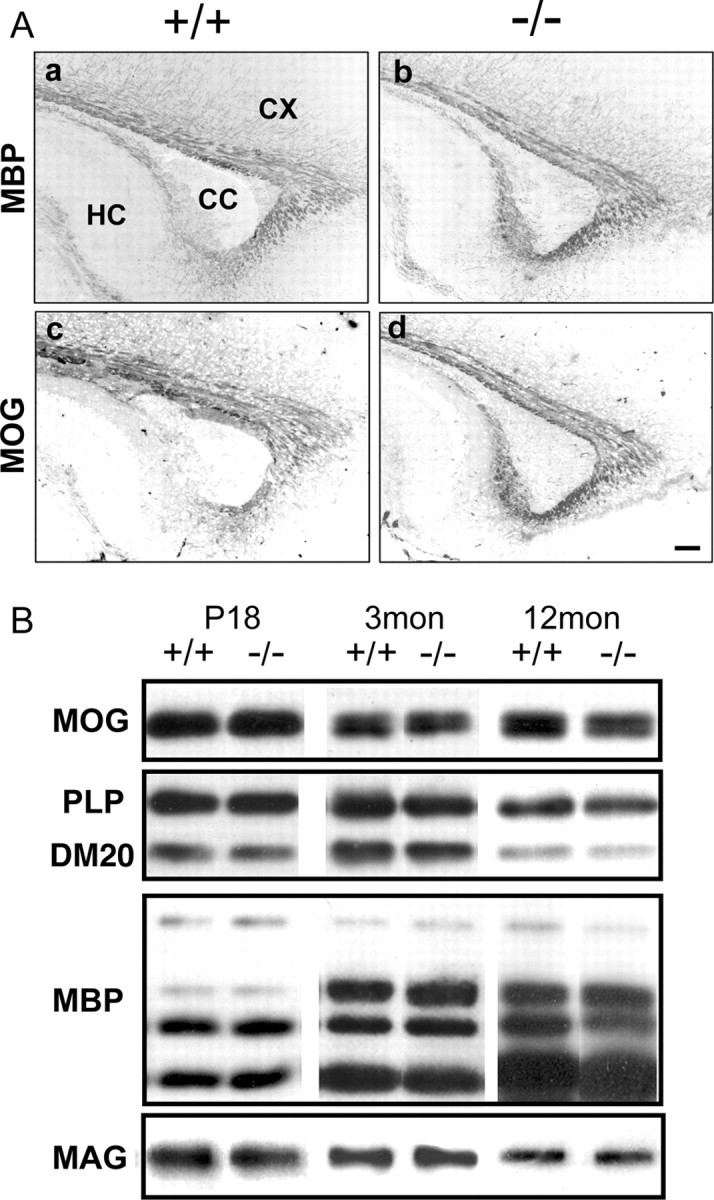
Myelination in Fgfr2 conditional knock-out mouse brain. A, Parallel parasagittal sections from P13 forebrain of control and FFC+ mice were analyzed by immunohistochemistry for MBP and MOG as markers of myelinated fibers. In the forebrain of FFC+ mouse (−/−), both MBP (a, b) and MOG (c, d) expressions appeared similar to control mice (+/+). Three to six sections each from two to four mice were analyzed from each group. CC, Corpus callosum; CX, cortex; HC, hippocampus. Scale bar, 100 μm. B, Immunoblot analysis of brain homogenates from control (+/+) and FFC+ (−/−) mice for the myelin proteins MOG, PLP/DM20, MBP, and MAG. No significant differences were observed between +/+ and −/− at 18 d, 3 months, or 12 months of age. A representative blot is shown.