
Figure 1.
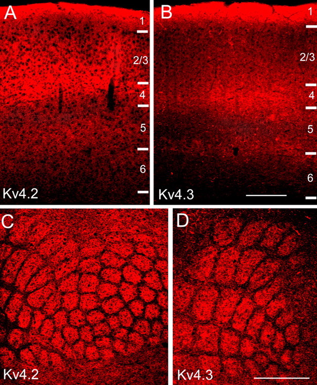
Expression levels of Kv4.2 and Kv4.3 in mouse primary visual and primary somatosensory cortex are layer specific. A, B, Immunofluorescence images of Kv4.2 and Kv4.3 expression in coronal sections through mouse primary visual cortex. Staining is densest in layers 1 and 4 and the layer 5/6 border. C, D, Kv4.2 and Kv4.3 immunofluorescence in tangential sections through mouse primary somatosensory cortex. Labeling is most intense in layer 4, in the center of barrels. Expression in septa between barrels is sparse. Scale bars: A, B, 0.2 mm; C, D, 1 mm.