
Figure 2.
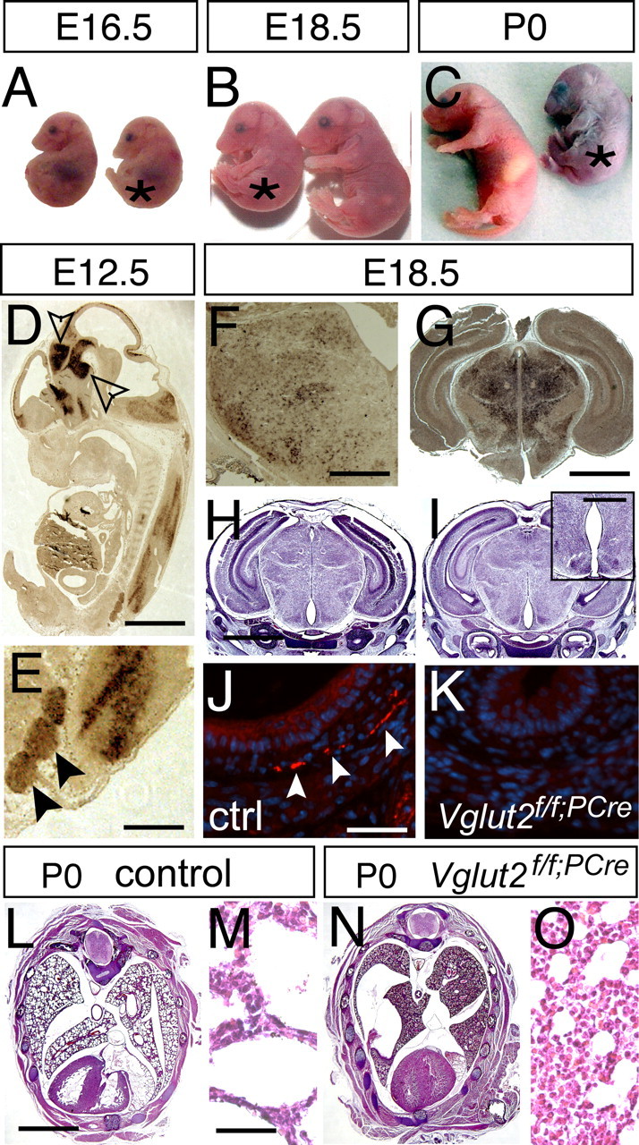
Vglut2 is expressed early in the embryo and is essential for normal development. A–C, Vglut2f/f;PCre embryos (*) appear arrested in development and can be distinguished from control littermates already at E16.5 by their hunched posture (A). At E18.5 (B), their smaller size (weights: control, 1.31 ± 0.03 g, n = 49; Vglut2f/f;PCre, 1.20 ± 0.03 g, n = 20; p = 0.0269) is easily distinguished at P0. C, The pups display cyanosis and die directly after birth. D, E, Vglut2 in situ hybridization of E12.5 embryos reveals strong mRNA expression in distinct brain regions (D), such as the ventral and dorsal diencephalon and ventral mesencephalon (arrows), brainstem, spinal cord, and dorsal root ganglia (close-up in E, marked with arrows) and also along the telencephalic and fourth ventricles and the aqueduct. F, G, Vglut2 continues to be strongly expressed in the brainstem (F) and the thalamus during late development (G). H, I, Hematoxylin/eosin staining of control (H) and Vglut2f/f;PCre embryos (I) coronal sections at E18.5 shows grossly normal brain histology of the null mutant compared with control, except a few animals (n = 4/9, E18.5 and P0) that displayed enlarged appearance of ventricles (e.g., third ventricle) (inset in I). J, K, VGLUT2-positive nerve terminals (red) are readily detected by immunofluorescence (nuclear costaining with DAPI in blue) in control internal organs at E18.5, as shown here in transverse sections of the muscular wall of the esophagus (J), whereas these nerve terminals are undetectable in the null mutants (K). L–O, Hematoxylin/eosin staining of transverse sections of control and Vglut2f/f;PCre P0 bodies shows grossly normal histology of the mutant compared with control except in the lungs, where the control display normal alveolar appearance (close-up in M), whereas an immature, noninflated structure is manifest in the mutant (close-up in O). Scale bars: D, 1 mm; E, 0.25 mm; F, 730 μm; G, 0.3 mm; H, 1.7 mm; I, inset, 0.5 mm; J, 45 μm; L, 2 mm; M, 40 μm.