
Figure 2.
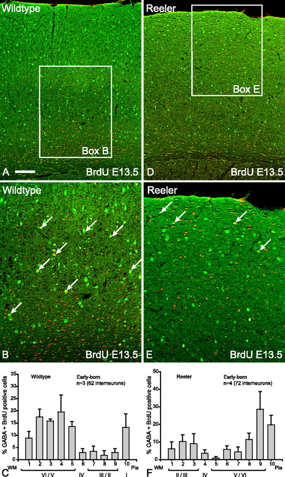
Early-born (E13.5) interneurons in reeler brains show layer inversion. A, Double- labeling for GABA (green) and BrdU (red) reveals that the vast majority of double-labeled cells (yellow) in wild-type cortex are in the lower layers. B, Higher magnification of boxed area in A indicating that the majority of double-labeled interneurons (arrows) are in the lower layers of the cortex. C, Histogram depicting the layer positions of GABA–BrdU interneurons (n = 3 animals, 62 cells; mean ± SEM percentage) indicating that the majority of early-born interneurons in wild-type cortices are distributed in the lower layers. D, In reeler cortex, double-labeled interneurons (yellow) born at E13.5 are found in the upper layers. E, Higher magnification of boxed area in D indicating that the majority of double-labeled (arrows) interneurons are in the upper layers of the cortex. F, Histogram depicting the layer positions of GABA–BrdU interneurons (n = 4 animals, 72 cells; mean ± SEM percentage) indicating that the majority of early-born interneurons in Reelin mutant cortices are distributed in the upper layers. Scale bar: A, D, 160 μm; B, E, 80 μm. WM, White matter.