
Figure 5.
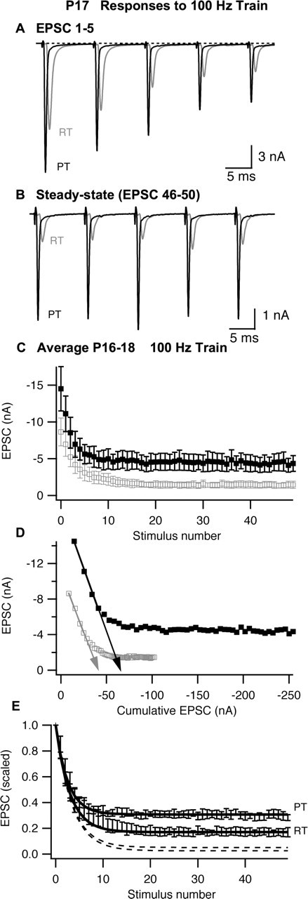
Synaptic depression recorded in P16–P18 MNTB neurons during stimulation of the calyx of Held with 100 Hz trains. A, B, The first five EPSCs (A) or last five EPSCs (B) recorded from a P17 MNTB neuron during a train of 50 stimuli at RT or PT. In B, stimulus artifacts were removed for clarity. C, Summary (n = 5 cells) of EPSC amplitudes. D, Determination of vesicle pool size and release probability by linear extrapolation (arrows) of EPSC amplitude versus cumulative EPSC amplitude. E, Normalized EPSC amplitudes. The dashed lines represent model predictions (see Materials and Methods) based on vesicle refilling rates obtained from measurements of recovery from synaptic depression (see Fig. 3) at RT or PT, and the solid lines represent model predictions when the recovery rate was adjusted to fit observed steady-state depression. EPSC amplitudes for each cell were scaled to the first EPSC in the respective train, and the mean and SEM of the normalized data set were calculated. In C and D, the open gray symbols represent data recorded at room temperature, and filled black symbols represent data recorded at physiological temperature; in E, symbols were omitted for clarity. Error bars represent SEM.