Figure 1.
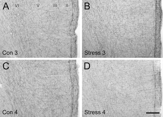
No obvious, consistent qualitative differences in the distribution, density, or general morphology of NET-ir fibers were observed in rats exposed to chronic cold stress. A–D, Representative light micrographs showing immunoperoxidase labeling of NET-ir axons in coronal sections through the rat PFC from the second cohort of control (Con) and chronically stressed (Stress) rats (animals 3 and 4). NET-ir fibers exhibit a uniform density across the cortical layers, with an additional dense plexus of axons coursing along the border between layers I and II. Scale bar, 250 μm.

