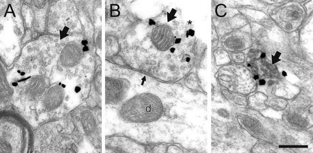
Figure 2.
NE axons and terminals (large black arrows) exhibit unusual immunoreactive characteristics in the PFC of control rats. A, B, By electron microscopic examination, most profiles express gold–silver labeling for NET predominantly in the cytoplasm and contain no detectable immunoperoxidase product for TH, even when they are cut open at the tissue surface where antibody penetration is maximal (asterisk in B). These profiles only occasionally form synapses, most commonly symmetric contacts (small black arrow in B) onto dendrites (d), and NET is not localized near these synapses. C, Only a minority of NE axons express NET predominantly on the plasma membrane, and these profiles also exhibit peroxidase labeling for TH. Scale bar, 0.48 μm.

