
Figure 8.
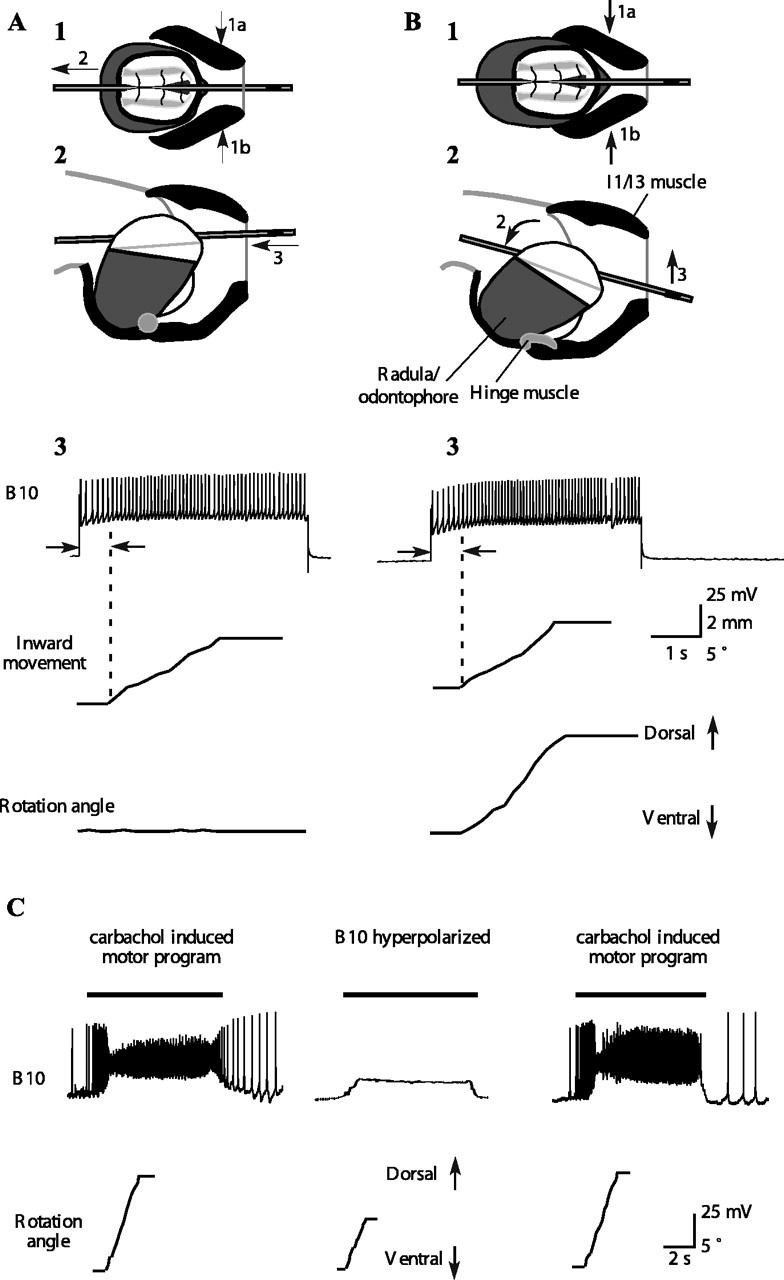
Activity in motor neuron B10 contributes to inward movement and dorsal rotation of a tube during ingestive-like feeding programs. A1, A2, Schematic top and side views of grasper and I1/I3/jaw complex in response to activating motor neuron B10 when the radula/odontophore is protracted to the peak position of a type A swallow. Direction of contraction of the I1/I3 muscle is shown by arrows 1a and 1b, and the resulting direction of the movement of the grasper is shown by arrow 2; in the side view (2), arrow 3 indicates the movement of the tube. A3, Activating B10 in a quiescent preparation protracted to the peak of a type A swallow induces a strong inward movement of a tube but no rotation. B1, B2, Schematic top and side views of grasper and I1/I3/jaw complex in response to activating motor neuron B10 when the radula/odontophore is protracted to the peak position of a type B swallow. Direction of contraction of the I1/I3 muscle is shown by arrows 1a and 1b, and the resulting direction of movement of the grasper is shown by arrow 2 in the side view; movement of the tube is indicated by arrow 3. B3, Activating B10 in a quiescent preparation protracted to the peak of a type B swallow induces a strong inward movement of the tube and a dorsal rotation. C, During carbachol-induced feeding motor program, B10 strongly depolarizes at the same time that the tube moves inward and rotates dorsally. Black bars above the B10 traces indicate the timing of the retraction phase. Hyperpolarizing B10 (middle) significantly reduces the inward movement and dorsal rotation. The magnitude of the inward movement and rotation is restored when B10 is no longer hyperpolarized (p < 0.05; n = 20 from 2 different animals; the inward movement of the tube was reduced from 4.4 ± 0.5 to 3.2 ± 0.7 mm).