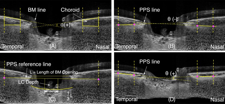
Figure 1.
Illustration of measurement of Bruch's membrane angle and of the peripapillary scleral angle. (A) Illustration of a v-shaped Bruch's membrane angle, α; (B) Illustration of an inverted-v shaped PPS angle, θ(−); (C) The measurement of the depth of anterior surface of the lamina cribrosa (LC depth) from the peripapillary sclera reference plane; (D) Illustration of a v-shaped PPS angle, θ(+).

