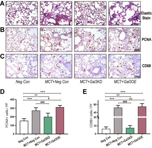
Figure 3. Gal-3 knockdown inhibits MCT induced vascular remodeling and Figure 3 Gal-3 knockdown suppress cell proliferation and macrophage infiltration.

(A-C) Elastic Van Gieson staining and immunostaining in lung tissue from Neg Con, MCT+ Neg Con, MCT+Gal3KD and MCT+Gal3OE group (A) Elastic Van Gieson staining staining to evaluate vasculature occlusive lesions and medial wall thickness of each group. (B) PCNA and (C) CD68 to assess proliferation of vessel cells and macrophages related inflammation separately. Arrow indicates representative PAs. (D and E) are semi-quantitative of PCNA (B) and CD68 (D) positive cells in high resolution fields of view (n=30), scale bars, 100μm. *** indicate P< 0.001, comparing Neg Con; ### indicate P<0.001, comparing MCT+Neg Con. Analyses performed by one-way ANOVA and Bonferroni post hoc. PF: per field.
