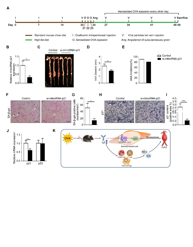
Figure 5. The effect of lincRNA-p21 on formation of AAA.
(A) Schematic diagram of animal model procedures. (B) Expression of lincRNA-p21 in AAA lesion of mice in control (n=8) and si-mlincRNA-p21 (n=5) groups. (C) Representative images of aortas from mice in control and si-mlincRNA-p21 groups of mice. (D) The average diameters of abdominal aortic aneurysm of two groups of mice. (E) The incidence rate of AAA in two groups of mice. (F) Representative pictures of β-galactosidase staining in AAA lesion. Scale bars: 25μm. (G) Senescent cells number per mm2 in AAA lesions. (H) Representative pictures of immunohistochemical staining for p21 in AAA lesions. Scale bars: 25μm. (I) The statistical analysis of p21 positive areas in immunohistochemical staining in AAA lesions. (J) RT-PCR detection of p21 and p53 mRNA expression in AAA lesions. (K) Schematic diagram of this article: IgE aggravates AAA mainly by upregulating lincRNA-p21 contributing to HSMC senescence. Data information: Data are presented as mean ± SEM. *P < 0.05; **P<0.01.

