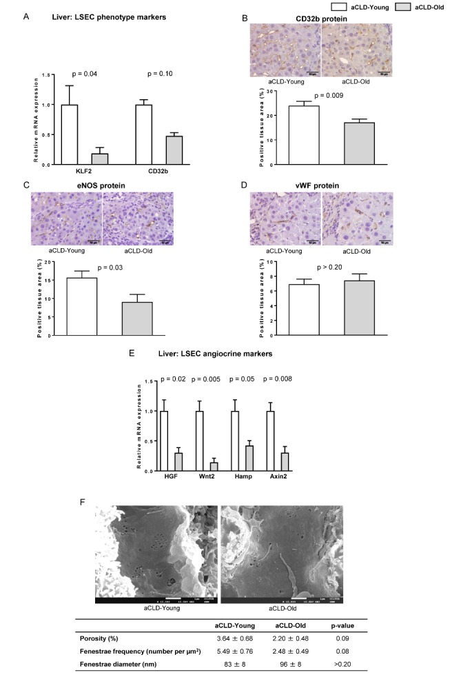
Figure 2. LSEC phenotype markers in aged rats with aCLD.

The following markers of sinusoidal endothelial phenotype were analysed in liver tissue from 4 months-young and 20 months-aged rats with aCLD. (A) mRNA expression of KLF2 and CD32b. (B) Representative images of CD32b immunehistochemistry and corresponding quantification. (C) Representative images of eNOS immunohistochemistry and corresponding quantification. (D) Representative images of vWF immunohistochemistry and corresponding quantification. (E) mRNA expression of HGF, Wnt2, Hamp and Axin2. (F) Representative scanning electron microscopy images & quantification of porosity, fenestration frequency and fenestration diameter. n=7 (A-E) and n=3 (F) per group. Results represent mean ± S.E.M. Images from B-D: 400X, scale bar=50μm. Images from F: 15000X, scale bar=1μm.
