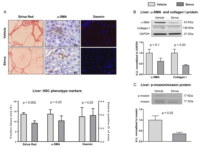
Figure 6. Simvastatin promotes decreased fibrosis deposition and HSC de-activation.
(A) Representative images of fibrotic content, α-SMA and desmin with their corresponding quantifications. (B) α-SMA and Collagen I protein expression in total liver tissue, normalized to GAPDH. (C) Representative western blots of moesin and p-moesin and corresponding quantification. n=10 (A-C). Results represent mean ± S.E.M. All images 400X, scale bar=50μm.

