
Figure 1. Expression and identification of phosphorylated RIPK1 in rat brains after ischemic injury.
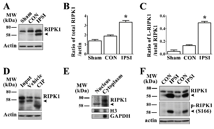
(A) Expression levels of RIPK1 in sham-operated (Sham) and ipsilateral (IPSI)/contralateral (CON) striatum at 24 h after MCAO. Expression levels of total RIPK1 (B) and low molecular weight RIPK1 (C) increased in the ipsilateral striatum compared with the contralateral striatum and sham-operated rats (n≥6). *p<0.05. (D) De-phosphorylation of RIPK1 in the ipsilateral striatum at 24 h after MCAO by calf intestinal alkaline phosphatase (CIP) treatment. (E) Expression levels of RIPK1 in the nuclear and cytoplasmic compartments of cells in the ipsilateral striatum, H3 was used as a specific nuclear marker and GAPDH as a specific cytoplasmic marker. (F) L-RIPK1 immuno-reacted with p-RIPK1 (Ser166) monoclonal antibody.