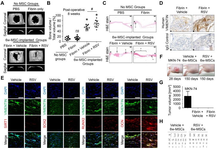
Figure 5. RSV induction improves bone healing potential of 6w-MSCs.
(A) Critical-sized calvarial defects (8-mm diameter) in rats were covered with fibrin glue, except for defect control treatment. Eight weeks after implantation, bone regeneration was measured by micro-computed tomography. A representative image is shown. (B) This graph shows the bone volume per mm3 (right panel) (n = 10). *, p < 0.05 compared to defect. #, p < 0.05 compared to 6w-MSCs treated with vehicle. (C) Hematoxylin and eosin staining was performed to observe new bone formation. The arrows show the edges of the host bone and line with asterisks indicates newly regenerated bone. Scale bar = 500 μm. (D) To confirm whether the newly regenerated bone was derived from a human origin, immunohistochemistry was performed using antibodies specific to human vimentin. The arrows indicate tissue derived from a human origin. Scale bar = 20 μm. (E) To confirm whether the transplanted 6w-MSCs contributed to bone regeneration of calvarial defects, immunohistochemistry was performed using antibodies against SIRT1, SOX2, RUNX2, and OCN as well as antibodies specific to human vimentin. The nucleus was stained with DAPI, and human VIMENTIN was stained with FITC-conjugated secondary antibody. SIRT1, SOX2, RUNX2, and OCN were stained with phycoerythrin (PE, red)-conjugated secondary antibody. Scale bar = 50 μm. (F) Effect of 6w-MSCs with RSV induction on tumorigenicity in 5-week-old female BALB/C nude mice. (G) Effect on the growth of MKN-74 cells, 6w-MSCs with vehicle induction, and 6w-MSCs with RSV induction xenografted in nude mice, showing no tumor growth in both 6w-MSC groups. (H) G-banding chromosome karyotype from 6w-MSCs with vehicle or RSV induction for 6 weeks. 6w-MSCs with 12th to 14th passages were used for animal experiments.

