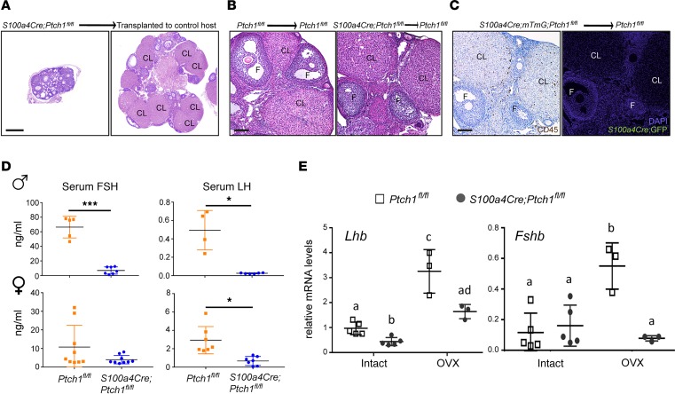
Figure 5. Gonad-extrinsic factors contribute to hypogonadism in Ptch1-mutant mice.
(A) Representative images of ovaries of S100a4-Cre;Ptch1fl/fl;mTmG mutant mice in situ (left) or transplanted to the bursa of control littermates (Ptch1fl/fl) for 4 weeks (right). CL, corpus luteum; F, follicle. Scale bar: 200 μm. (B) Representative images of ovaries of Ptch1fl/fl control mice (left) and S100a4-Cre;Ptch1fl/fl;mTmG mutant mice (right) transplanted to control littermates (Ptch1fl/fl) for 4 weeks. Scale bar: 100 μm. (C) Representative images of IHC staining of CD45 and IF staining of GFP on adjacent serial sections of ovaries from S100a4-Cre;Ptch1fl/fl;mTmG mutant mice transplanted to Ptch1fl/fl control mice. Scale bar: 100 μm. (D) Serum concentration of FSH and LH in control and homozygous Ptch1 mutant male and female mice at 8 weeks of age. *P < 0.05; ***P < 0.001; Student’s t test. (E) Relative mRNA levels of pituitary Lhb and Fshb in wild-type controls and homozygous Ptch1 mutants with and without ovariectomy (OVX, n = 3 for samples from OVX mice; n = 5 for samples from intact mice). Total RNA was assayed by qPCR and the concentration of each transcript was normalized to that of housekeeping gene Rpl19. Data are represented as mean ± SD. Bars without common letters are significantly different (P < 0.05); two-way ANOVA. All data are from mice at 8 weeks of age.

