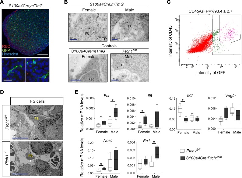
Figure 7. Expression of S100a4-Cre is restricted to CD45+ cells, including FS cells, in the anterior pituitary and leads to malfunction of these cells.
(A) Representative images of IF staining for GFP in the anterior pituitary of S100a4-Cre;mTmG reporter control mice. RBC, red blood cell. Scale bars: 200 μm and 20 μm. (B) Representative images of transmission electron microscopy (TEM) of immunogold labeling of GFP on pituitary tissue sections from S100a4-Cre;mTmG reporter mice at 8 weeks of age. Arrows point to immunogold signals. FS, folliculo-stellate cell; EC, endothelial cell. Scale bars: 200 nm (top panels) and 500 nm (bottom panels). (C) The percentage of CD45-positive cells among GFP-positive cells in the pituitaries of S100a4-Cre;mTmG reporter control mice analyzed by flow cytometry. The image represents results from 4 independent samples. (D) Representative images of TEM of FS cells in control and Ptch1-mutant mice. FS cells are identified according to their ultrastructural features. Scale bar: 10 μm. (E) Relative mRNA levels of genes involved in the pituitary microenvironment in whole pituitary tissues of wild-type controls and homozygous Ptch1 mutants at 8 weeks of age (n ≥ 5). Total RNA was assayed by qPCR and the concentration of each transcript was normalized to that of the housekeeping gene Rpl19. Data are represented as mean ± SD. *P < 0.05; Student’s t test. All data are from mice at 8 weeks of age.

