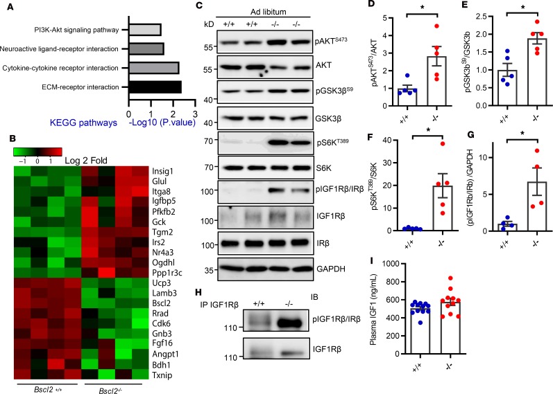
Figure 1. Elevated basal IGF1R-mediated PI3K/AKT signaling in hypertrophic Bscl2–/– hearts.
(A and B) Transcriptome and pathway analyses of differentially expressed (DE) genes and heatmap from DE genes related to PI3K/AKT signaling in left ventricles of 10-week-old female Bscl2–/– mice after a 4-hour fast (n = 4 pooled from 3 animals each). (C–G) Western blot and quantification of pAKT at Ser473, pGSK3β at Ser9, pS6K at Thr389, and pIGF1Rβ/IRβ at Tyr1158/Tyr1162/Tyr1163 in hearts (n = 5/group). (H) Immunoprecipitation of cardiac IGF1Rβ detects enhanced tyrosine phosphorylation of IGF1Rβ. Representative Western blot is shown. n = 5/group. (I) Plasma IGF1 levels (n = 11/group). For C–I, ad libitum–fed 3-month-old male Bsc2+/+ and Bscl2–/– mice were used. *P < 0.05 by unpaired t test.

