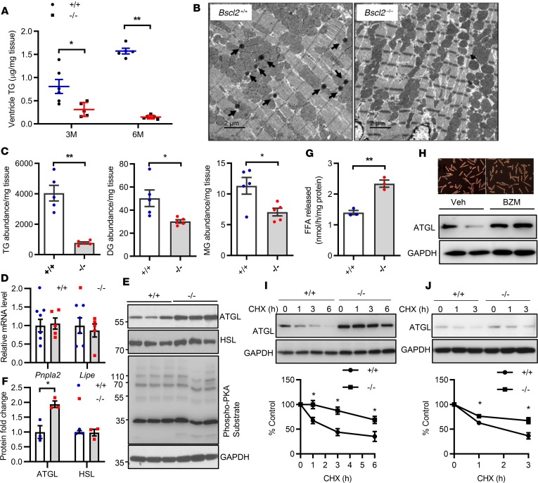
Figure 2. BSCL2 deficiency reduces intramyocellular glycerolipids and elevates ATGL stability and expression in the heart.
(A) Quantitative enzymatic analyses of ventricle triglyceride (TG) in 3- and 6-month-old (3M and 6M) nonfasting Bscl2+/+ (+/+) and Bscl2–/– (–/–) mice (male, n = 4–7/group). (B) Representative transmission electron microscopy of 6-month-old nonfasting male Bscl2+/+ and Bscl2–/– hearts. Arrows indicate lipid droplets. Scale bars: 2 μm. (C) Comparison of the total normalized ion abundances for glycerolipids including TG, diacylglycerol (DG), and monoacylglycerol (MG) identified by lipidomics in hearts of 6-month-old male Bscl2–/– mice fed ad libitum (n = 5 with each pooled from 3 animals). (D) RT-PCR analyses of Pnpla2 and Lipe gene expression in hearts of 3-month-old nonfasting mice (n = 5–7/group). (E and F) Representative Western blot and quantification of heart protein expression in 3-month-old male Bscl2+/+ and Bscl2–/– mice fed ad libitum (n = 3/group). (G) TG hydrolase activity in 3-month-old heart homogenates incubated with radiolabeled 3H-triolein. Free fatty acid (FFA) release was measured and normalized to protein (male, n = 3 in triplicate). (H) Viability and ATGL expression in primary adult mouse cardiomyocytes isolated from male C57BL/6J mice after incubation with vehicle (Veh) or 100 nM bortezomib (BZM) for 12 hours. (I and J) Cycloheximide (CHX) shutoff analysis of endogenous ATGL turnover in primary adult mouse cardiomyocytes isolated from 3-month-old male Bscl2+/+ and Bscl2–/– mice and in Bscl2+/+ and Bscl2–/– MEFs. Densitometry from Western blots was standardized to ATGL expression at 0 hours. *P < 0.05; **P < 0.005 by unpaired t test (C and G) or multiple t tests after correction using the Holm-Sidak method (A, D, F, I, and J).

