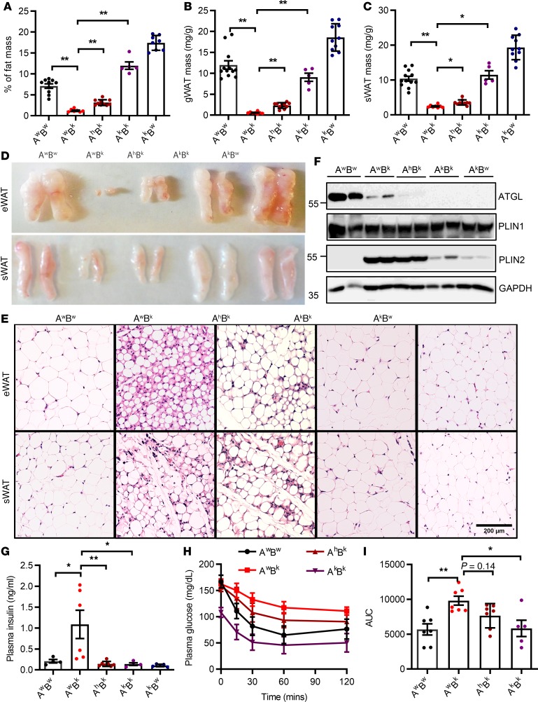
Figure 4. ATGL ablation rescues lipodystrophy and its associated insulin resistance in Bscl2–/– mice.
(A–C) Percentage of fat mass assessed by NMR, and masses of gonadal white adipose tissue (gWAT) and subcutaneous WAT (sWAT) as normalized to body weight. (D and E) Representative images and H&E staining of gWAT and sWAT. Scale bar: 200 μm. (F) Representative Western blotting of mature adipose marker and lipid droplet protein (LDP). (G) Plasma insulin levels after 4-hour fast. For A–G, 10-week-old female Atgl+/+Bscl2+/+ (AwBw), Atgl+/+Bscl2–/– (AwBk), Atgl+/–Bscl2–/– (AhBk), Atgl–/–Bscl2–/– (AkBk), and Atgl–/–Bscl2+/+ (AkBw) littermates were used for all experiments (n = 5–10/group). Limited numbers of male AkBk mice were obtained. However, a similar extent of rescue of lipodystrophy was observed in male AhBk and AkBk mice. (H and I) Insulin tolerance test and area under the curve (AUC) in mice other than AkBw (male and female, n = 5–7/group). *P < 0.05; **P < 0.005 by 1-way ANOVA with Dunnett’s multiple-comparisons correction.

