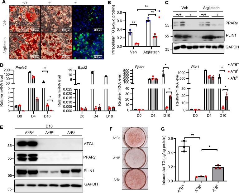
Figure 5. ATGL inhibition partially rescues adipocyte differentiation of Bscl2–/– cells.
Bscl2+/+ and Bscl2–/– MEFs were subjected to a standard hormone cocktail DMI (dexamethasone, IBMX, and insulin) to induce adipocyte differentiation. Four days (D4) after differentiation, cells were treated with vehicle (Veh) or 10 μM Atglistatin and kept until D10. (A) Oil Red O and HCS LipidTOX Green neutral lipid staining. Scale bars: 200 μm. (B) Intracellular triglyceride (TG) content (2-way ANOVA with post hoc Tukey’s test) and (C) representative Western blot of PPARγ and PLIN1 at D10 after DMI induction. (D–G) Stromal vascular cells isolated from Atgl+/+Bscl2+/+ (AwBw), Atgl+/+Bscl2–/– (AwBk), and Atgl–/– Bscl2–/– (AkBk) mice were subjected to DMI-induced adipocyte differentiation. (D) mRNA expression of Pnpla2, Bscl2, Pparγ, and Plin1 was measured at D0, D4, and D10 after DMI induction (2-way ANOVA with Dunnett’s multiple-comparisons post-hoc correction). (E) Representative protein expression, (F) Oil Red O staining, and (G) intracellular TG content at D10 after adipocyte induction (1-way ANOVA with Dunnett’s correction for multiple comparisons). *P < 0.05; **P < 0.005.

