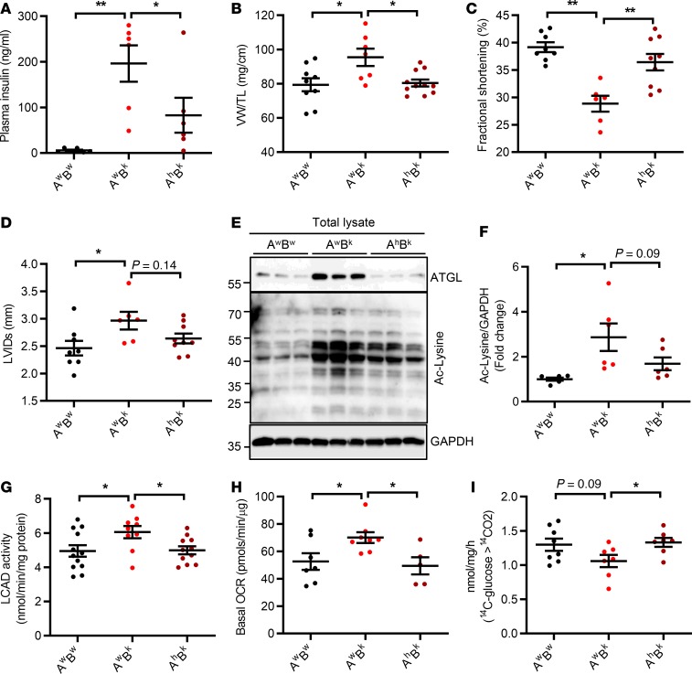
Figure 6. Partial ATGL inactivation rescues hypertrophy and cardiomyopathy in Bscl2–/– mice.
(A) Nonfasting plasma insulin levels, (B) ventricle weight (VW) normalized to tibia length (TL), and (C and D) fractional shortening (%) and left ventricle internal diameter at systole (LVIDs, mm). (E and F) Representative Western blotting (E) and fold changes of acetylated lysine (Ac-Lysine) as normalized to GAPDH (F) in whole heart. (G) Cardiac LCAD activity, (H) basal oxygen consumption rate (OCR) in isolated cardiac mitochondria, (I) CO2 production after incubating heart crude mitochondrial fraction with 14C-glucose. Six-month-old Atgl+/+Bscl2+/+ (AwBw), Atgl+/+Bscl2–/– (AwBk), and Atgl+/– Bscl2–/– (AhBk) male mice were used, n = 5–12/group. *P < 0.05, **P < 0.005 by one-way ANOVA with Dunnett’s multiple-comparisons correction.

