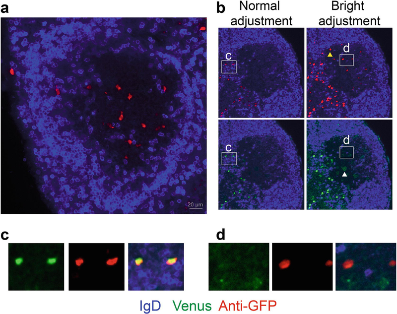
Fig. 3.
Representative microscopy of the Verigem reporter in LN cryostat sections. Heterozygous Verigem reporter mice were immunized subcutaneously with NP-KLH in alum adjuvant, and LNs were collected 7 d later. (a) Example of a GC (IgDlow region) with IgE+ B cells detected by anti-GFP staining (red). (b) Demonstration of the difference in sensitivity between anti-GFP staining and Venus fluorescence and the importance of the display adjustments to detect IgE+ PCs (Venushigh) versus IgE+ GC B cells (Venuslow). All four panels show the same image with either anti-GFP staining (red, upper panels) or with Venus fluorescence (green, lower panels). The image display levels are adjusted in a standard fashion to show the full range of signal detected (left panels) versus an exaggerated narrow range making the red/green signal appear bright (right panels). Note the GC B cells (e.g., see the yellow arrowhead) can only be detected by anti-GFP staining on the right panels with the bright adjustment of display levels. While PCs can be directly detected by either Venus fluorescence or anti-GFP staining (inset c) with normal display levels, the GC B cells cannot be visualized by Venus fluorescence but can be visualized by the anti-GFP staining with the bright adjustment of display levels (inset d). Within the GC, the bright display adjustment of the Venus channel instead reveals autofluorescent cells, such as tingible body macrophages (e.g., see the white arrowhead in (b, lower right panel)). The bright adjustment of display levels also results in oversaturation of the PCs; thus it may be difficult to properly visualize PCs and GC B cells with the same display settings. Anti-GFP was detected in the far red channel (see Note 10)

