Figure 1.
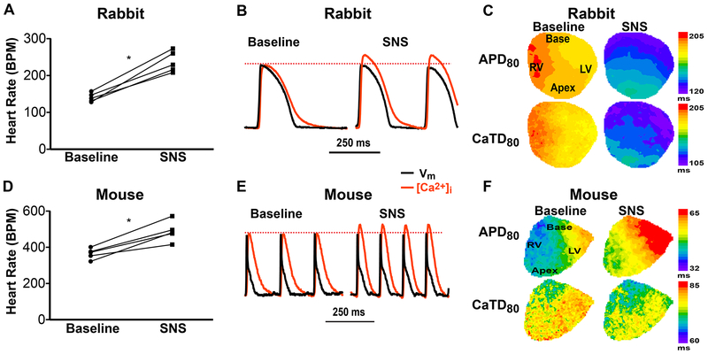
Effects of sympathetic nerve stimulation (SNS, 20 sec) in innervated isolated rabbit and mouse hearts. (A) SNS increased rabbit heart rate (*p<0.05 SNS vs. baseline, n=5). (B) Example optical action potentials (APs) and Ca2+ transients (CaTs) at baseline and during SNS showing obvious shortening of AP duration (APD), CaT duration (CaTD) and an increase in CaT amplitude (when normalized to baseline amplitude) in the rabbit heart. (C) Example maps of APD and CaTD at baseline and during SNS in the rabbit heart. (D) SNS increased mouse heart rate (*p<0.05 SNS vs. baseline, n=5). (E) Example optical traces as in (B) from a mouse heart. (F) Example maps of APD and CaTD at baseline and during SNS in the mouse heart.

