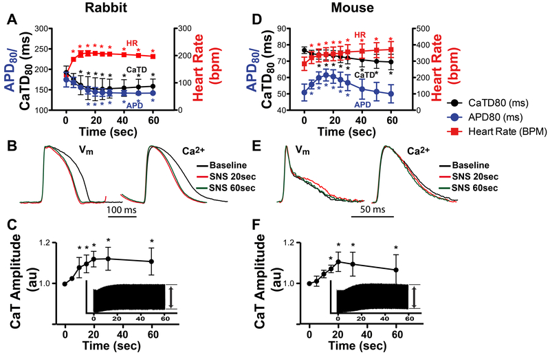
Figure 2.
Effects of sympathetic nerve stimulation (SNS, 60 sec) on heart rate (HR), action potential duration (APD) and Ca2+ transients (CaT). (A) In rabbit, APD and CaT duration (CaTD) monotonically decreased and HR monotonically increased over 60 sec of stimulation (*p<0.05 vs. time 0, n=4). (B) Example optical APs and normalized CaTs at baseline and at 20 and 60 sec of stimulation. (C) Relative rabbit CaT amplitude during SNS normalized to baseline (time 0). Inset shows continuous CaT recording over 60 sec from which relative amplitudes were calculated (*p<0.05 vs. time 0, n=6). (D) In mouse, HR monotonically increased, CaTD monotonically decreased, and APD displayed a biphasic response (*p<0.05 vs. time 0, n=5). (E) Example mouse optical APs and CaTs showing APD prolongation at 20 sec SNS. (F) Relative mouse CaT amplitude during SNS (*p<0.05 vs. time 0, n=6).

