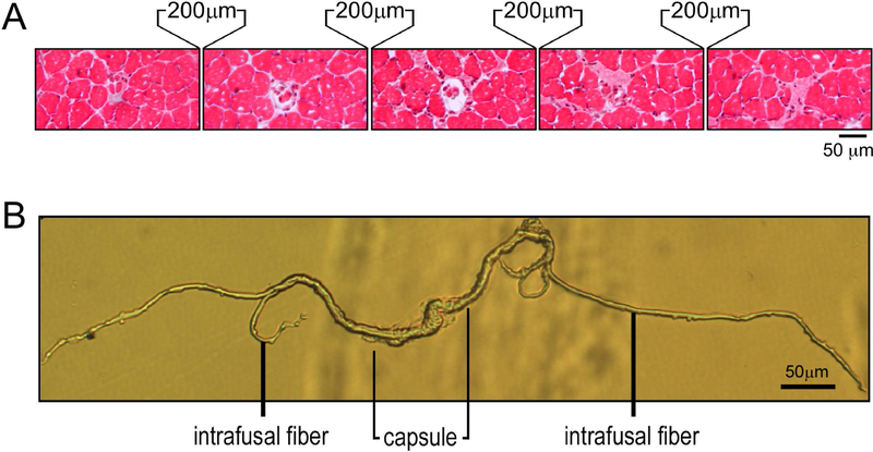
Figure 1. Identification and isolation of mouse muscle spindles.
A. Serial cross sections of a mouse soleus muscle at 200 µm intervals revealed both the central capsule and intrafusal myofibers of a muscle spindle. B. A muscle spindle isolated from mouse soleus muscle using enzymatic digestion and micro-dissection showing the central capsule and intrafusal myofibers.

