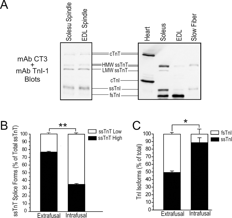
Figure 2. Intrafusal fibers have different thin myofilament protein contents from that in extrafusal fibers.
A. The representative TnT and TnI Western blots in the left panel show that ssTnT is expressed at significant levels in spindle intrafusal fibers of WT mouse with the low molecular weight splice form predominant. cTnT is also expressed in adult mouse muscle spindle intrafusal fibers. Slow skeletal muscle TnI was the major TnI isoform in muscle spindles. Total protein extracts from adult mouse cardiac, slow and fast muscles are shown as controls in the right panel. B & C. Densitometry quantification of the Western blots demonstrate the significantly different ssTnT splice form and TnI isoform contents of isolated intrafusal fibers and total muscle protein extracts representing extrafusal fibers. *P<0.05; **P<0.001.

