Figure 1.

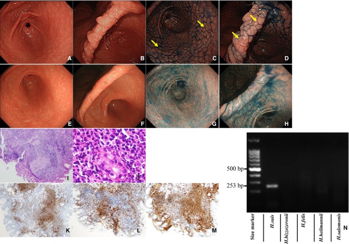
Characterization of case A1. Endoscopic examination (before eradication): Multiple heterogeneous nodules similar to nodular gastritis in antrum (A) and anglus (B). Indigo carmine dye makes the nodules more obvious in the antrum (C) and anglus (D). Endoscopic examination (after eradication): no nodules in the antrum (E) or anglus (F), even with indigo carmine dye in the antrum (G) and anglus (H). Pathological examination (before eradication): Lymphoma cells proliferate diffusely and form heterogeneous nodules with lymphoid hyperplasia. HE, ×100 (I). Atypical lymphoid cells invading the epithelium, resulting in the destruction of mucosal glands and formation of lymphoepithelial lesions. HE, ×400 (J). Immunohistochemistry (before eradication): Neoplastic cells are positive for CD20 (K), negative for CD3 (L), and positive for CD79a (M). PCR assay (before eradication): Positive for Helicobacter suis (253 bp); negative for other non‐H pylori Helicobacter (N). Biopsies taken from areas indicated with yellow arrows showed pathological findings compatible with mucosa‐associated lymphoid tissue lymphoma. Abbreviation: HE, hematoxylin and eosin
