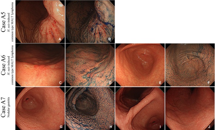
Figure 5.

Characterization of cases A5, A6, and A7. Cases A5 and A6: Endoscopic pictures of common type MALT lymphoma with Helicobacter suis infection. In case A5, the superficial phenotype of gastric MALT lymphoma was observed in the gastric body under white light (A) and chromoendoscopy (B). In case A6, the superficial phenotype was observed in the gastric body under white light (C) and chromoendoscopy (D). Mild nodular gastritis without MALT lymphoma was observed in the gastric antrum under white light (E) and chromoendoscopy (F). Case A7: Representative endoscopic pictures of nodular gastritis. Endoscopic examination (before eradication): Multiple homogeneous nodules are seen in the antrum. Whitish spots exist on the top of each nodule (G). No nodule is observed in the gastric anglus (H). Mild atrophy is observed in the gastric body (I). Indigo carmine dye makes the nodules more obvious in the antrum (J). Abbreviation: MALT, mucosa‐associated lymphoid tissue
