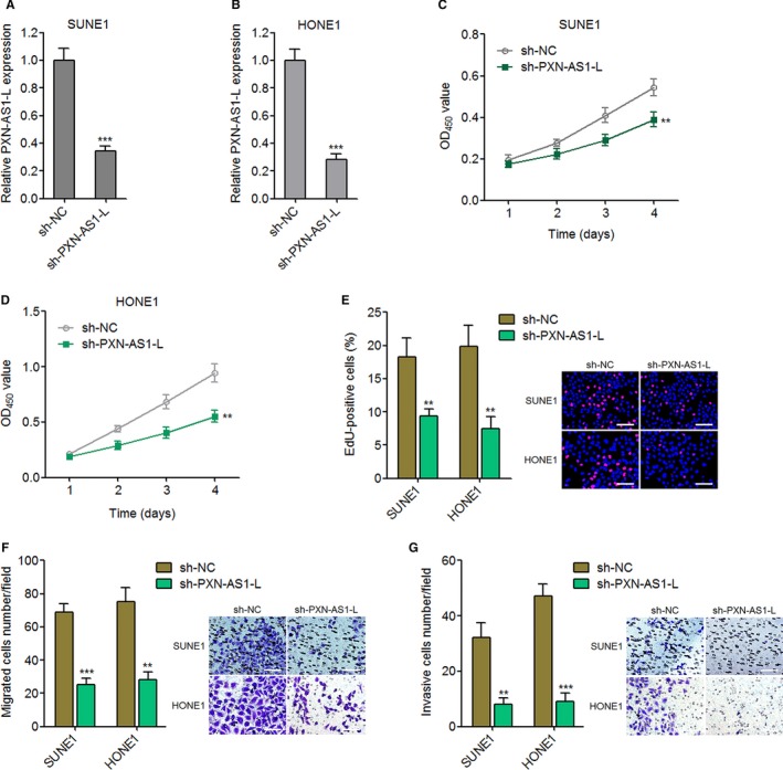
Figure 3.

Silencing of PXN‐AS1‐L suppresses nasopharyngeal carcinoma cell proliferation, migration, and invasion. A, PXN‐AS1‐L expression levels in PXN‐AS1‐L stably silenced and control SUNE1 cells were determined by qPCR. B, PXN‐AS1‐L expression levels in PXN‐AS1‐L stably silenced and control HONE1 cells were determined by qPCR. C, Cell proliferation of PXN‐AS1‐L stably silenced and control SUNE1 cells was determined by Cell Counting Kit‐8 (CCK‐8) assay. D, Cell proliferation of PXN‐AS1‐L stably silenced and control HONE1 cells was determined by CCK‐8 assay. E, Cell proliferation of PXN‐AS1‐L stably silenced and control SUNE1 and HONE1 cells was determined by ethynyl deoxyuridine (EdU) incorporation assay. Scale bars, 100 μm. F, Cell migration of PXN‐AS1‐L stably silenced and control SUNE1 and HONE1 cells was determined by transwell migration assay. Scale bars, 100 μm. G, Cell invasion of PXN‐AS1‐L stably silenced and control SUNE1 and HONE1 cells was determined by transwell invasion assay. Scale bars, 100 μm. Results are displayed as mean ± SD from 3 independent experiments. **P < 0.01, ***P < 0.001 by Student's t test
