Figure 5.

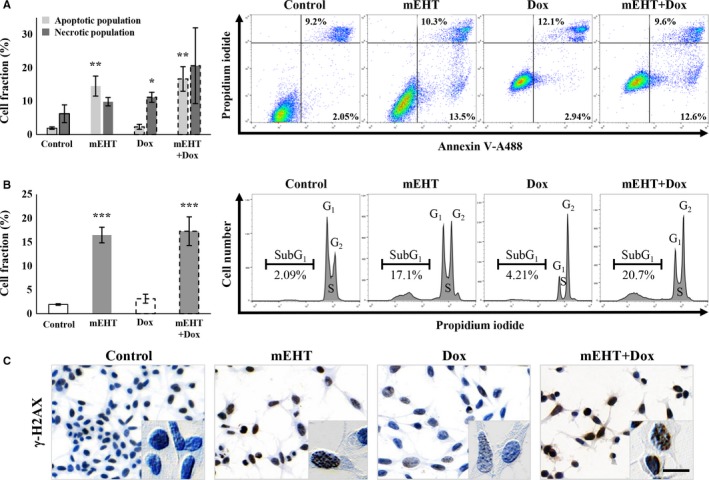
Comparison of the ratio of cell and DNA damage 24 h after treating cultured C26 tumor cells. Significantly elevated apoptotic cell fractions after mEHT and necrotic cell fractions after Dox treatments and their additive, merged effect after combination (mEHT + Dox) therapy (A). Significant increase in subG1 phase cell fractions both after mEHT and combined treatments refer to the apoptosis‐related DNA damage (B), where DNA double‐strand breaks were indicated by upregulated γ‐H2AX granular positivity (brown) in cell nuclei using immunocytochemistry (C). Scale bar: 100 µmol/L. *P < 0.05; **P < 0.01; ***P < 0.001
