
Fig. 1.
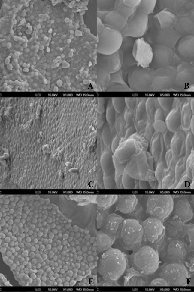
Representative images of S. cerevisiae cells acquired by SEM with increase of 1000 and 5000 times: a, b natural yeast (NY); c, d mechanically ruptured yeast (MRY) and e, f modified autolysis yeast (MAY)
Official websites use .gov
A
.gov website belongs to an official
government organization in the United States.
Secure .gov websites use HTTPS
A lock (
) or https:// means you've safely
connected to the .gov website. Share sensitive
information only on official, secure websites.
Representative images of S. cerevisiae cells acquired by SEM with increase of 1000 and 5000 times: a, b natural yeast (NY); c, d mechanically ruptured yeast (MRY) and e, f modified autolysis yeast (MAY)