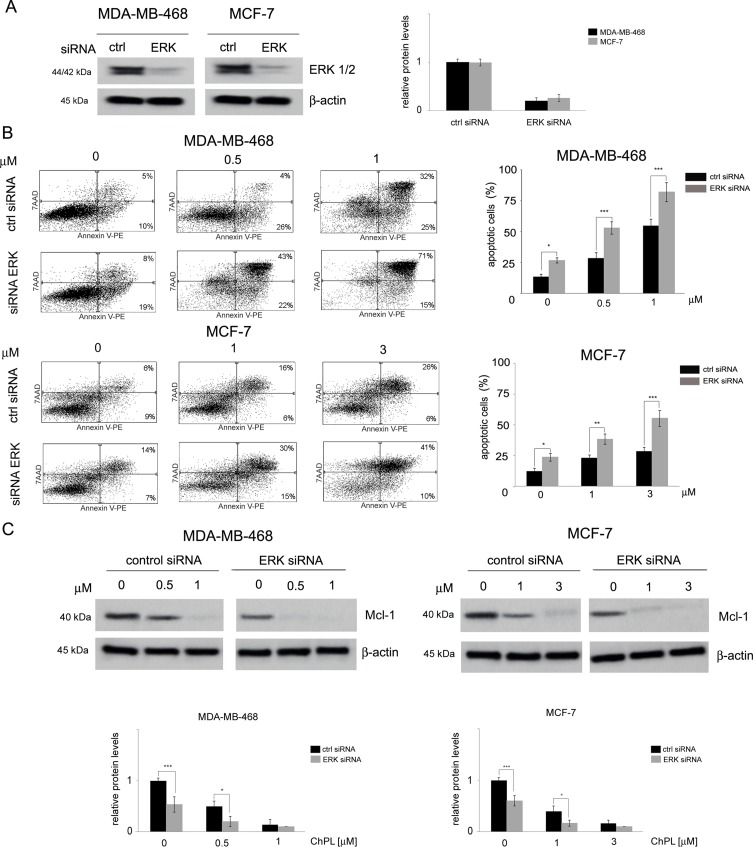
Figure 5.
The role of MAPK signaling in 3-chloroplumbagin-mediated ERK inhibition and apoptosis induction. (A) Silencing of ERK1/2 with ERK1/2 siRNA and control siRNA in MDA-MB-468 and MCF-7 cells. Protein levels were determined with Western blot analysis. Densitometric analysis represents ERK1/2 levels normalized to β–actin levels. (B) Apoptosis induction by ChPL in MDA-MB-468 and MCF-7 cells transfected with ERK siRNA or control siRNA. Apoptosis induction was assessed with flow cytometry after Annexin V-PE staining. (C) Influence of ERK silencing on expression levels of Mcl-1 in MDA-MB-468 and MCF-7 cells treated with ChPL. Mcl-1 levels were assessed with Western blot analysis. Densitometric analysis represents Mcl-1 levels normalized to β-actin levels. Data were analyzed by one-way ANOVA with Tukey’s post hoc tests [p < 0.05 (*), p < 0.01 (**), p < 0.001 (***), n = 3].

