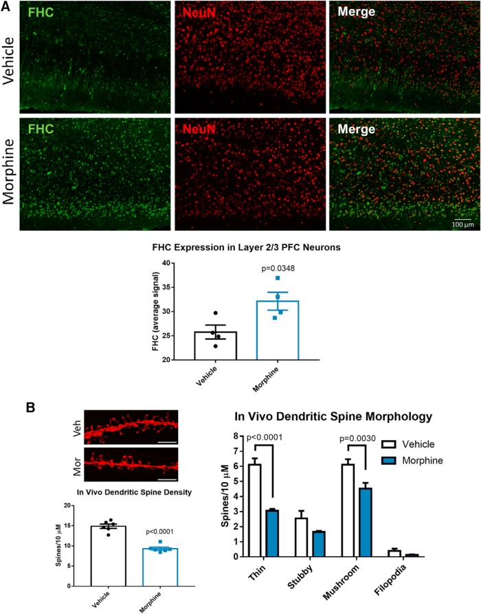
Figure 4.
Morphine upregulates FHC and decreases mature dendritic spines in layer 2/3 neurons of the rat medial prefrontal cortex. A, Morphine upregulates FHC in cortical neurons in vivo. Three-week-old Holtzman rats were treated with extended-release morphine pellets (25 mg) or placebo for 96 h as detailed in the methods, followed by perfusion and brain tissue collection. Brain sections were stained with antibodies against FHC (green) and the neuronal marker NeuN (red), and images were acquired with a 20× objective. Images were analyzed by measuring the staining intensity of FHC in NeuN-positive areas of the layer 2/3 prelimbic cortex of the mPFC. FHC staining intensity values from individual neurons were averaged to one value per rat, represented as one dot in the graph. FHC staining was significantly higher in neurons of morphine-treated rats; N = 4 rats per treatment group. Data analyzed by Student’s t test; t(6) = 2.717. B, Morphine reduced thin and mushroom dendritic spine density in PFC neurons. A different group of three-week-old Holtzman rats treated with morphine or placebo pellets were used for dendritic spine analysis. PFC-containing tissue slices were stained with DiI to visualize dendritic spines, as shown in the micrograph; scale bar = 5 µm. Morphine decreased the overall spine density of layer 2/3 prelimbic cortex neurons (t(10) = 8.482), and specifically reduced the density of thin and mushroom spines. Stubby spines and filopodia were not significantly changed by morphine; N = 6 rats per treatment group. Spine density data were analyzed by Student’s t test, and morphology data were analyzed by two-way ANOVA with Sidak’s multiple comparisons test (treatment F(1,40) = 44.5, p < 0.0001; morphology F(3,40) = 114, p < 0.0001).

