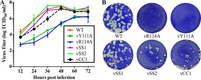
FIG 9.
In vitro growth characterization of SHFV wild-type (WT) and mutant viruses. (A) Multiple-step virus growth curves of WT and mutant viruses. Each data point shown represents the mean value from duplicated treatments. Error bars show standard errors of the means (SEM). (B) Plaque morphologies of WT SHFV and mutants thereof. Confluent cell monolayers were infected with 10-fold serial dilutions of the virus suspension. After 2 h of incubation, an agar overlay was added on top of the infected cells. Plaques were observed after 3 days of incubation at 37°C. Cells were stained with 0.1% crystal violet.

