FIG 1.
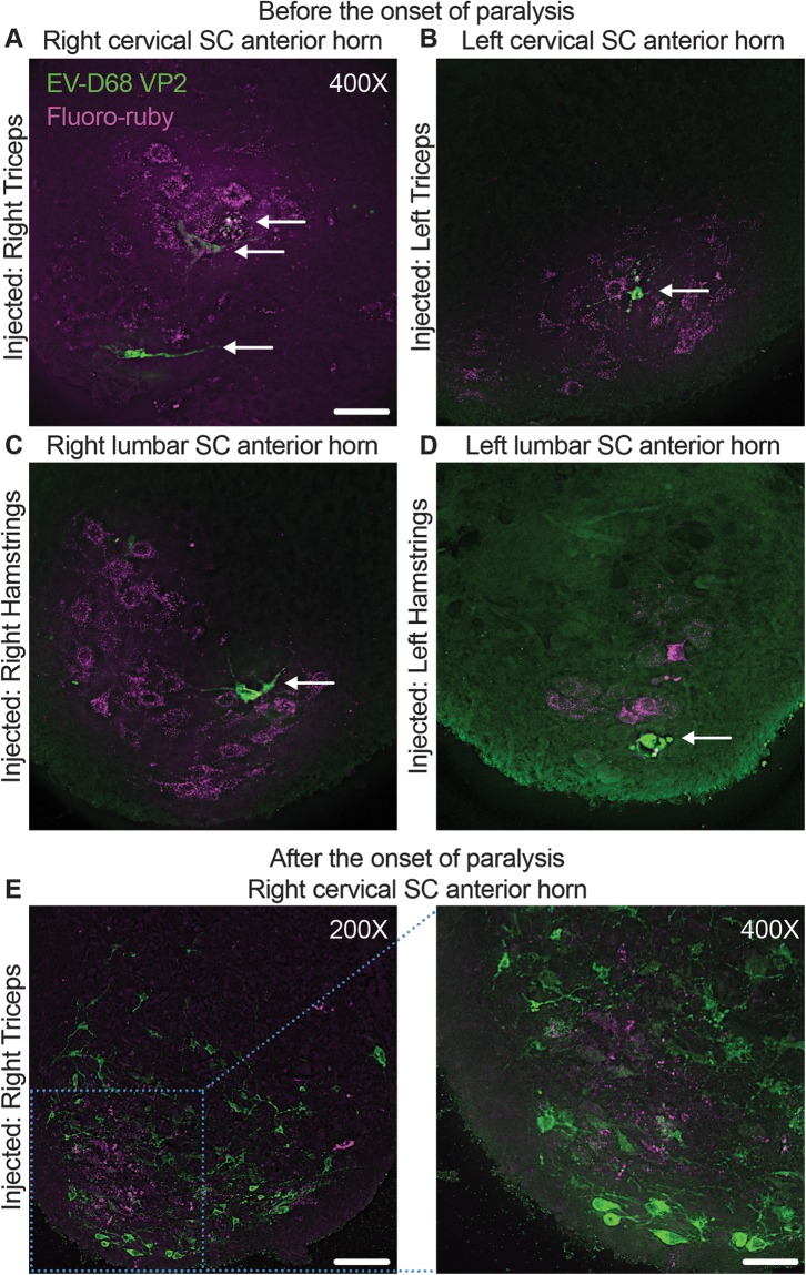
Retrograde axonal transport of EV-D68 in the mouse spinal cord. Mice were coinjected with 10% fluoro-ruby and 1,000 TCID50 of EV-D68 IL/14-18952 on postnatal day 2 (P2) of life into either the right triceps (A), left triceps (B), right hamstring (C), or left hamstring (D) muscle and examined for staining of EV-D68 capsid protein VP2 (arrows) before the onset of paralysis on day postinfection (dpi) 2 or 3. The first detectable EV-D68 antigen appeared in motor neurons in the spinal cord (SC) anterior horns corresponding to the injected muscle, as indicated by the presence of fluoro-ruby tracer dye in the right cervical spinal cord anterior horn (A), left cervical spinal cord anterior horn (B), right lumbar spinal cord anterior horn (C), and left lumbar spinal cord anterior horn (D). (E) A mouse injected with 1,000 TCID50 EV-D68 IL/14-18952 and 10% fluoro-ruby into the right triceps muscle on P2 began showing signs of moderate paralysis on dpi 3. Images from the cervical spinal cord anterior horn show the death and loss of the fluoro-ruby-expressing neurons and spread of EV-D68 to the surrounding neurons. Bars = 100 μm for the ×200-magnification image and 50 μm for the ×400-magnification images.

