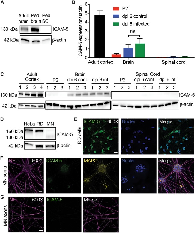
FIG 6.
Characterization of ICAM-5 expression in humans, mice, and motor neurons. (A) ICAM-5 expression by Western blotting (WB) in the human adult temporal cortex (adult brain) compared to expression in the pediatric (Ped) frontal cortex (Ped brain) and cervical spinal cord (Ped SC) from a 3-year-old human child. ICAM-5 was not detected in the pediatric cervical spinal cord. All samples contained 10 μg of total protein. (B) Neonatal mice were evaluated for ICAM-5 RNA expression normalized to β-actin expression by RT-qPCR on P2 (the standard age of injection; n = 3) as well as on dpi 6 following i.m. injection of 1,000 TCID50 of EV-D68 IL/14-18952 (n = 3) or the control used for mock injection (n = 3). Cortical tissue from 8-week-old mice (n = 4) was used as a positive control. There was no significant difference between ICAM-5 RNA expression on day postinfection 6 for the control and EV-D68-injected mouse brains (P = 0.5386), as evaluated by one-way ANOVA between all groups followed by Tukey’s multiple-comparison test. (C) ICAM-5 expression by Western blotting mirrored the findings of RT-qPCR in the adult mouse cortex (n = 4) as well as P2 (n = 3), dpi 6 infected (n = 3), and dpi 6 control (cont; n = 3) brains and spinal cords. All samples contained 20 μg of total protein. (D) ICAM-5 protein expression in RD and HeLa cells and iPSC motor neurons (MN) by Western blotting. All samples contained 20 μg of total protein. (E) The strong ICAM-5 protein expression seen in RD cells was confirmed by immunohistochemistry (IHC). (F and G) Faint, punctate ICAM-5 staining was observed in the iPSC motor neuron soma and dendrites (F) by IHC but not in the axons (G). Images represent results from three experimental replicates. Bars = 20 μm.

