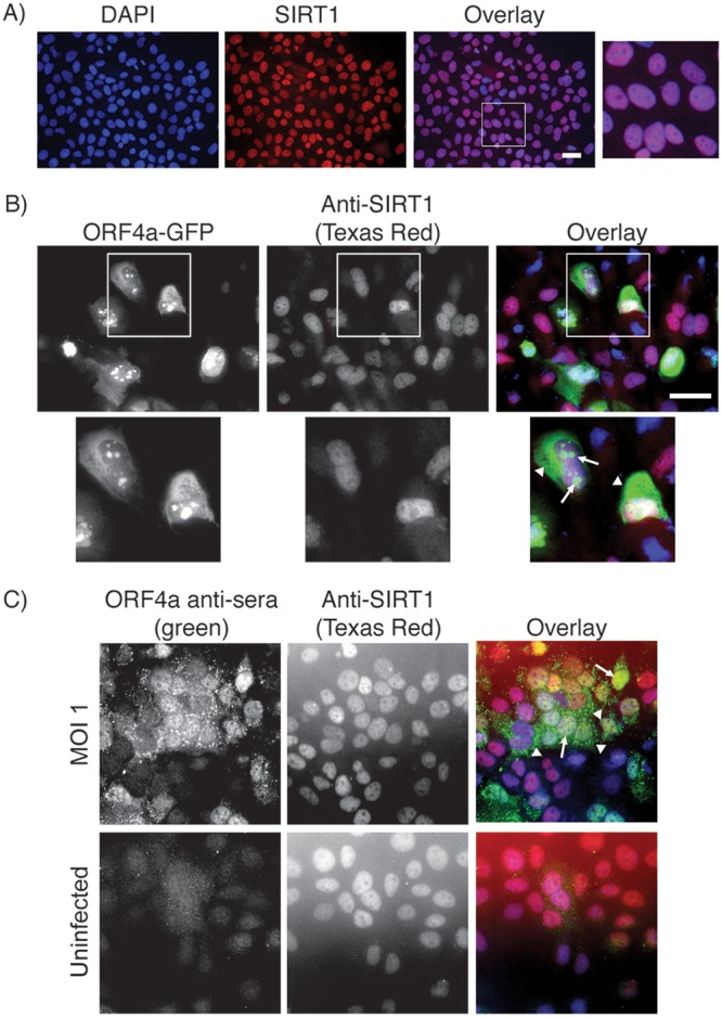
FIG 2.

Localization of ORF4a and SIRT1 in Huh7 cells. (A) Huh7 cells grown on coverslips were subjected to immunofluorescence staining with anti-SIRT1 antibody (visualized with anti-mouse Texas Red). (B) Huh7 cells grown on coverslips were transfected with a plasmid expressing ORF4a-GFP and were subsequently subjected to immunofluorescence labeling for SIRT1 (visualized with anti-mouse Texas Red). Arrows point toward nuclear and arrowheads point toward cytosolic ORF4a localizations. (C) Huh7 cells were plated to coverslips 1 day prior to infection with MERS-CoV at an MOI of 1 (uninfected cells were grown as a control). Infection was allowed to proceed for 18 h prior to fixation and immunofluorescence labeling for SIRT1 (visualized with anti-mouse Texas Red) and ORF4a (visualized with anti-rabbit FITC). Arrows point toward nuclear and arrowheads point toward cytosolic ORF4a localizations. In all experiments, nuclei were labeled with DAPI. Scale bars = 50 μm.
