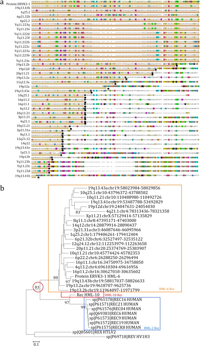
FIG 6.
Multiple alignment and phylogenetic relationships of HML-6 Rec domains. (a) Multiple alignment of the HML-6 Rec amino acid sequences with the protein ERVK3-1 used as reference. The colors in the sequences show disagreements in the alignments; black lines represent the deletion. ORFs are indicated by orange arrows, eventually stopping in correspondence of stop codons (black dots) or frameshift mutation (black arrows). (b) Relationship between the four best-preserved HML-6 Rec domains and the known HML-2 and HML-10 Rec domains is shown in a phylogenetic tree. This relationship was ascertained by using the NJ method and the Kimura two-parameter model with 1,000 bootstrap replicates.

